
Overview of the Circulatory System
The circulatory system consists of the heart and blood vessels; hence it is also called the cardiovascular system, where cardio pertains to the heart and vascular pertains to the blood vessels.
Blood vessels are composed of arteries, veins, and capillaries. There are two types of circulation: the pulmonary circulation and the systemic circulation. Pulmonary circulation is responsible for oxygenation of the blood that will be transported throughout the body via systemic circulation [1].
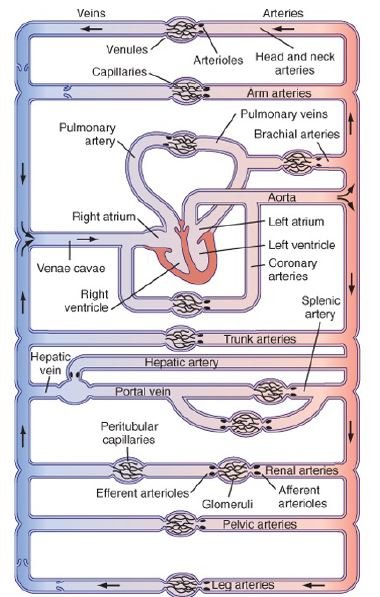
Picture 1: Schematic Diagram of the Circulatory System
Source: Koeppen BM & Stanton BA. Berne & Levy Physiology 6th edition. Elsevier, Inc. 2008.
In the pulmonary circulation, deoxygenated blood from the peripheral veins drain to the superior and inferior venae cavae towards the right atrium and right ventricle. Deoxygenated blood is then pumped to the lungs via pulmonary arteries. The lungs will get rid of the wastes and reoxygenate the blood. Oxygenated blood will pass through the pulmonary veins into the left atrium and left ventricle then back into the systemic circulation [1-3].
The heart pumps the oxygenated blood and circulates it to all parts of the body through the arteries. Arteries serve as the hydraulic filter because the continuous blood flow pumped by the heart is stabilized before it approaches the capillaries. Capillaries serve as the exchange point between oxygenated and deoxygenated blood. When it reaches the target organs, the target organs use up the oxygen that the blood carries, so it becomes deoxygenated blood. Veins carry deoxygenated blood back into the heart for reoxygenation and the cycle continues [1-5].
This article focuses on the differences between the blood vessels.
Layers of the Blood Vessels
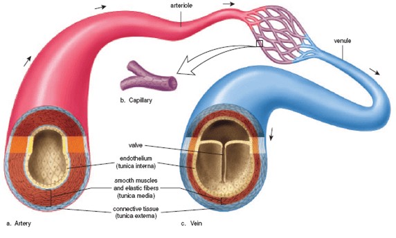
Picture 2 : Layers of the Blood Vessels
Source: majordifferences.com
The layers of the blood vessels basically determine their function. Both arteries and veins have tunica interna, tunica media, and tunica externa.
Tunica interna is the innermost layer made up of endothelial cells and is in contact with the blood. Capillaries only have this single layer of endothelial cells which makes it possible for diffusion of oxygen, carbon dioxide, nutrients, and waste products to take place.
Tunica media is the middle layer composed of smooth muscles and elastic fibers which regulates blood flow and pressure by vasoconstriction and vasodilation. This layer is thicker in arteries compared to veins that is why blood flow and pressure in the arteries are higher than in the veins.
Tunica externa is the outermost layer composed of connective tissue that serves as support to the entire blood vessel. This layer is also thicker in arteries that the veins to accommodate the higher blood flow and pressure [1,6,7].
Description of the Blood Vessels: Arteries vs. Veins vs. Capillaries
The blood vessel responsible for distributing oxygenated blood throughout the body is the artery. Since the blood is oxygenated, its color is bright red and when an artery gets injured, blood squirts because of arterial pressure is high compared to the vein [5,8].
The blood vessel that comes out from the heart is the aorta, the largest artery in the body. As the arteries continue peripherally, the wall becomes thinner, the internal diameter narrows, and they tend to be more muscular until they become arterioles. Since the surface area in the aorta and large arteries are relatively broad, blood pressure is relatively high, and resistance to blood flow are relatively low compared to smaller peripheral arteries and arterioles where the surface area is narrow so there is lower pressure and higher resistance to blood flow [7,9].
From the arterioles, the capillary beds (each consisting of 10-100 capillaries) arise. This is where the exchange of oxygen, nutrients, and wastes between blood and tissues take place through diffusion. This is made possible in the capillaries by its very thin walls, highly permeable endothelial cells, very short tubes, and low flow velocity [7,10].
From the capillaries, venules arise. Its side become bigger until they become veins. Veins are responsible for bringing deoxygenated blood back to the heart for reoxygenation so the body can use the blood further. As it approaches the heart, the veins become fewer in number, cross-sectional area becomes decreased, and the blood flow velocity increases. Also, veins have valves that prevent backflow of blood towards the organs, especially in the lower extremities, due to the effects of gravity. Arteries do not have valves [1,5,7,9].
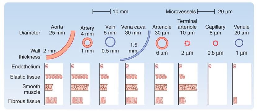
Picture 3: Measurement of blood vessels in terms of diameter, wall thickness, endothelium, elastic tissue, smooth muscle, and fibrous tissue
Source: Koeppen BM & Stanton BA. Berne & Levy Physiology 6th edition. Elsevier, Inc. 2008.
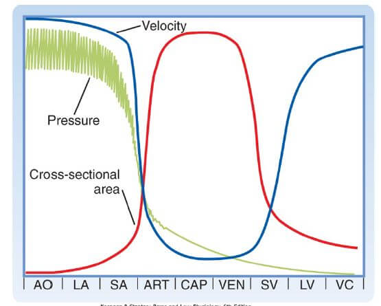
Picture 4: Relationship between blood pressure, blood flow velocity, and cross-sectional area of the blood vessel. Decreased cross-sectional area means higher pressure and velocity, as seen in large blood vessels like aorta, large arteries, large veins, and venae cavae. Increased cross-sectional area means lower pressure and velocity, as seen in arterioles, capillaries, and venules.
Source: Koeppen BM & Stanton BA. Berne & Levy Physiology 6th edition. Elsevier, Inc. 2008.
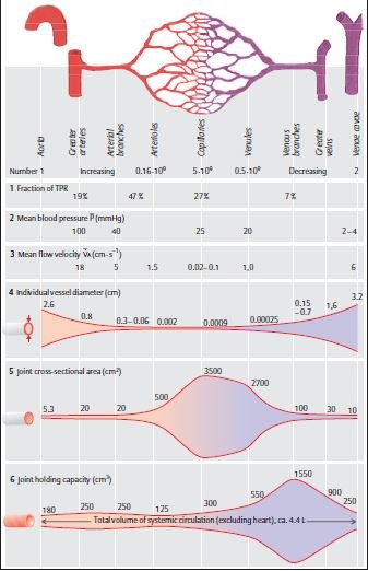
Picture 5: Characteristics of Blood Vessel Segments
Source: Despopoulos A & Silbernagl S. Color Atlas of Physiology 5th edition. Thieme. 2003.
Comparison Chart
| VARIABLE | ARTERY | VEIN |
| Internal Diameter | Narrower (4mm) | Wider (5mm) |
| Wall Thickness | Thicker (1mm) | Thinner (0.5mm) |
| Strength | More | Less |
| Distensibility | Less | More |
| Smooth Muscle | More | Less |
| Elastic Tissue | More | Less |
| Fibrous Tissue | More | Less |
| Cross-Sectional Area* | Decreased | Decreased |
| Pressure* | High | Low |
| Volume | Low (15%) | High (65%) |
| Flow Velocity* | HIghest | Low |
| Direction of Blood Flow | Away from the heart | Towards the heart |
| Oxygen in the Blood | Oxygenated blood (except for pulmonary artery which carries oxygenated blood) | Deoxygenated blood (except for pulmonary vein which carries oxygenated blood) |
| Color | Bright red | Dark red |
| Valves | Absent | Present |
| Injury to the Blood Vessel | Squirting blood | Pooling of blood |
*Depends on the size of the blood vessel. There is inverse relationship between cross-sectional area vs. pressure and flow velocity.
Videos
- This video shows the simple anatomy of arteries and veins. Watch it on: https://www.youtube.com/watch?v=ByB7cK9iTKA
- This video emphasizes the main differences between the arteries and veins. Watch it on https://www.youtube.com/watch?v=7b6LRebCgb4
References
- http://www.diffen.com/difference/Arteries_vs_Veins
- http://www.differencebtw.com/difference-between-artery-and-vein/
- https://www.sharecare.com/health/circulatory-system-health/what-difference-between-an-artery-and-a-vein
- Despopoulos A & Silbernagl S. Color Atlas of Physiology 5th edition. Thieme. 2003.
- http://www.majordifferences.com/2013/02/difference-between-artery-and-vein.html#.WE9dxlN97IU
- https://www.reference.com/science/difference-between-veins-arteries-capillaries-981c90258bf1ba22
- https://www.boundless.com/biology/textbooks/boundless-biology-textbook/the-circulatory-system-40/mammalian-heart-and-blood-vessels-226/arteries-veins-and-capillaries-853-12098/
- http://www.mananatomy.com/basic-anatomy/difference-arteries-veins
- Koeppen BM & Stanton BA. Berne & Levy Physiology 6th edition. Mosby Elsevier, Inc. 2008.
- Guyton & Hall Textbook of Medical Physiology 12th edition. Saunders Elsevier, Inc.