Introduction
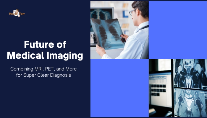
Medical imaging has already revolutionized healthcare—from X-rays unveiling broken bones to MRI scanning deep into soft tissues.
Yet further innovation looms: hybrid imaging fusing MRI, PET (positron emission tomography), CT, and other techniques into a single platform or integrated workflow.
Such combinations—like PET-MRI or SPECT-CT—promise richer data with less patient inconvenience, enabling earlier disease detection and precision therapy planning
. This article dives into the potential of next-generation multi-modal imaging, the science that underlies it, and the ways it might transform diagnoses from neurology to oncology.
The Rationale Behind Multimodal Imaging
Limitations of Single Modalities
Each imaging technique has strengths:
- MRI excels at soft-tissue contrast, clarifying structures in the brain or joints.
- PET reveals metabolic or molecular activity—for instance, highlighting active tumors.
- CT quickly shows anatomy or bone detail.
However, by themselves, they may overlook complementary data. Merging modalities yields a full picture: structural, functional, and molecular.
Synergy for Early Detection
PET’s sensitivity to minute biochemical changes can detect tumors sooner, but it might lack anatomical precision. Combined with MRI or CT, doctors localize suspicious hot spots exactly. For complex conditions (like certain neurological or cardiac diseases), a multi-layered approach clarifies subtle pathologies early, potentially guiding targeted interventions.
Cutting-Edge Modalities
PET-MRI
PET-MRI merges PET’s functional/metabolic imaging with MRI’s high-resolution soft tissue. This synergy is particularly valuable in:
- Oncology: Precisely localizing active cancer regions and analyzing infiltration of surrounding tissues.
- Neurology: Identifying specific brain metabolism changes correlated with structural changes (e.g. in Alzheimer’s or epilepsy).
- Cardiology: Evaluating myocardium viability while visualizing structural anomalies.
SPECT-CT
SPECT (Single Photon Emission Computed Tomography) combined with CT is widely used in nuclear medicine—for example, in bone scans or cardiac perfusion. CT provides an anatomical map onto which SPECT data overlays, clarifying lesion location and bone or organ detail.
Future: PET-CT-MRI Tri-Modality?
Some advanced research centers explore triple combos or universal machines that can pivot between MRI sequences, CT scanning, and PET acquisitions. Though expensive and complex, they might significantly reduce total scanning time and yield unprecedented integrated datasets.
AI and Data Fusion
Merging Data Sets
Combining MRI, PET, CT images yields massive volumes of data. AI helps align and fuse them into coherent 3D visualizations. Radiologists or surgeons might see color-coded functional overlays (PET) on top of crisp anatomical references (MRI/CT), guiding highly accurate diagnoses or planning.
Automated Insight
Machine learning can compare multi-modal scans to massive reference databases, flagging suspicious patterns or subtle anomalies that even expert eyes might miss. Over time, AI might also estimate prognosis or predict therapeutic responses from integrated imaging data.
Personalized Treatment
As precision medicine grows, integrated imaging plus AI can tailor treatments—for example, identifying which part of a tumor is more metabolically active (from PET) and precisely shaping radiotherapy fields accordingly, while MRI guides structural margins.
Applications in Various Fields
Oncology
Multi-modal imaging is a cornerstone of modern cancer staging and therapy monitoring. PET or SPECT highlight tumor metabolism, while MRI/CT show infiltration or structural relationships. Post-treatment scans track remission or detect recurrence early.
Neurology
For epilepsy surgery, combined PET (locating hyperactive foci) and MRI (pinpointing structural anomalies) produce more accurate resection maps.
In stroke or degenerative disorders, multi-modal imaging can differentiate salvageable tissue from irreversibly damaged areas, guiding interventions.
Cardiology
Cardiac PET-CT or SPECT-CT images both perfusion and anatomical structure—helpful in diagnosing coronary artery disease or viability of myocardium post-infarction.
Advanced MRI sequences measure tissue elasticity or microvascular flow, giving deeper insights into heart function.
Patient and Clinical Benefits
One-Stop Imaging
Combined machines reduce multiple visits. A single session captures structural, functional, and metabolic data. This synergy shortens diagnostic timelines, reducing overall radiation exposure if the patient would otherwise require separate scans.
More Accurate Diagnosis
Integrating data from multiple imaging types lowers false positives or ambiguous interpretations. For example, a suspicious mass might show up on PET but then be confirmed as benign upon seeing MRI’s detailed soft-tissue structure.
Personalized Therapy Planning
Detailed imaging fosters targeted treatments—like precisely shaped radiotherapy fields sparing healthy tissue, or planning minimal surgical routes. Minimally invasive interventions or advanced chemo regimens can rely on robust multi-modal guidance.
Cost, Complexity, and Other Hurdles
Expensive Equipment
PET-MRI scanners cost significantly more than stand-alone MRI or PET. Not all hospitals can afford them. Additionally, operational costs and specialized staff training are substantial, limiting widespread adoption.
Radiation and Time
Although integrated approaches can reduce separate scanning, some protocols may still require extended scanning durations. CT or PET exposures add up. Striking a balance between thoroughness and minimal radiation is crucial.
Data Overload
Multi-modal imaging yields enormous data sets. Radiologists must manage huge files, ensuring robust IT infrastructure and advanced post-processing software. Without efficient data handling and AI-driven assistance, scanning could outpace reading capacity.
The Road Ahead
[H3] Smaller, Cheaper Hybrid Machines
As research refines hardware, we might see more compact, cost-effective integrated systems—like PET-MRI “lite” for smaller centers. If advanced manufacturing lowers the price point, adoption grows, democratizing multi-modal imaging.
AI’s Expanding Role
AI might handle real-time “scan adaptation”—where scanning parameters adjust mid-exam based on initial results. Or it automatically merges images, creating 4D modeling for dynamic processes (like beating hearts or moving joints). This synergy yields deeper clinical insights.
Molecular Imaging Breakthroughs
Beyond classic PET tracers (like FDG), next-generation tracers may target specific proteins or gene expressions. Combining them with structural scans yields precision diagnosis for early cancers or metabolic dysfunction, ushering in truly personalized medicine.
Advice for Patients and Providers
- Ask About Multi-Modal Options: If dealing with complex diagnoses, see if a center offers PET-MRI or SPECT-CT—some conditions significantly benefit from integrated imaging.
- Balancing Benefit and Cost: For common tasks (e.g., a routine check), single-modality might suffice. For complicated or uncertain cases, the added detail of multi-modal can be invaluable.
- Check Insurance Coverage: Advanced imaging is expensive. Confirm coverage or out-of-pocket costs, especially for specialized multi-modal tests.
- Stay Current: Imaging technology evolves quickly. Providers should seek continuing education on new protocols, while patients can monitor updates for newly recommended screening or follow-ups.
Conclusion
Multi-modal imaging—combining MRI, PET, CT, and potentially other technologies—offers a glimpse of the future in diagnostic clarity. By uniting each modality’s strengths, clinicians gain unprecedented detail on structure,
function, and molecular activity—all in a single session. While cost and complexity currently limit widespread rollout
, the relentless progress in hardware miniaturization, AI data fusion, and cost reduction signals that tomorrow’s standard approach to scanning complex diseases may be an all-in-one synergy. For patients
, this means more accurate, timely, and personalized diagnoses—and, ultimately, better outcomes in the battles against cancer, heart disease, neurological conditions, and beyond.
References
- Pichler BJ, Wehrl HF, Judenhofer MS. Latest advances in molecular imaging instrumentation. J Nucl Med. 2008;49(Suppl 2):5S–23S.
- Zaidi H, del Guerra R. An outlook on future design of hybrid PET/MRI systems. Med Phys. 2011;38(10):5667–5689.
- Townsend DW. Multifunctional PET/CT imaging in oncology. Eur J Nucl Med Mol Imaging. 2004;31(Suppl 1):S131–S143.
- Cherry SR, Louie AY, Jacobs RE. The integration of multimodality imaging in biomedical research. Nat Rev Mol Cell Biol. 2008;9(7):669–676.
- LaForest R, et al. PET/MRI in oncology: current status and perspectives. Curr Radiol Rep. 2013;1:187–196.
- Cerami C, et al. The role of PET-MRI in neurodegenerative conditions. Semin Nucl Med. 2020;50(4):338–348.
- Bartolozzi C, Donati F. Molecular imaging in oncology: changing the concept of tumor detection. Radiol Med. 2015;120(10):914–923.
- Gambhir SS. Molecular imaging of cancer with integrated MRI–PET modalities. Nat Rev Cancer. 2020;20(6):365–380.
- Bailey DL, Townsend DW, Valk PE, Maisey MN. Positron Emission Tomography: Basic Science and Clinical Practice. Springer; 2005.
- Maurer AH. SPECT/CT imaging in general nuclear medicine. Nucl Med Commun. 2012;33(4):349–357.


