
Definition: What is the Central Sulcus of Rolando?
A sulcus is a defined as a deep groove that separates the gyri or convolutions on the surface of the brain.
Named after Luigi Rolando, the central sulcus (of Rolando) or Rolandic fissure is a part of the brain that extends from the cerebral longitudinal fissure up to near the lateral sulcus (of Sylvius) or Sylvian fissure perpendicularly.
It is one of the most prominent fissures in the brain. It separates the frontal and parietal lobes of the brain [1, 2, 3].
Embryologically, the central sulcus appears at around 5th to 6th months of fetal life. It ascends towards the superior margin of the cerebral hemisphere and becomes definitive at the 8th month. It is usually a continuous sulcus but according to Retzius and Eberstaller, an interrupted sulcus is found in 1% of cases.
The space in the superior end of the central sulcus is called crochet Rolandique or crochet de Rolando. The inferior terminals of the precentral and postcentral gyri are connected by a bridge called opercule Rolandique or pli de passage frontoparietal inferieur [4].
Paul Broca was the first one to describe the superior and inferior ends of the central sulcus. A deeper fissure within it exists and he called it the pli de passage frontoparietal moyen (PPFM) or the deep annectant gyrus by Cunningham. A study confirmed that a localized elevation of this is the anatomical hallmark of the hand representation [5].
Anatomy and its Pictures: Where is it Located?
A layman would think that the grooves or fissures that he sees in the brain have one name. But advanced neuroanatomy would tell that each of these have their own names and functions. If we look at the brain, it appears to be too complex. Well, yes it is. It is too complex it is hard for a layman, and sometimes even for physicians, to identify which is which. But once you get to know the clues, you can identify the names of the neuroanatomical parts quite easily.
The following pictures will guide you in determining the anatomical position of the central sulcus of Rolando.
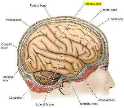
Picture 1 Lateral View of the Brain within the Skull
Image Source: Snell RS, Clinical Neuroanatomy 7th edition, Lippincott Williams & Wilkins 2010
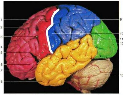
Picture 2 : The central sulcus of Rolando is shaded in white. It divides the frontal lobe (red) and parietal lobe (blue).
Image Source: Rohen JW et al, Color Atlas of Anatomy 7th edition, Lippincott Williams & Wilkins 2011

Picture 3 Lateral View of the Cerebral Hemisphere
Image Source: Haines DE, Fundamental Neuroscience 6th edition

Picture 4 : Medial View of the Cerebral Hemisphere
Image Source: Haines DE, Fundamental Neuroscience 6th edition

Picture 5 : Sagittal Section of the Brain in Situ
Image Source: Netter FH et al, Atlas of Neuroanatomy and Neurophysiology, Icon Custom Communications 2002

Picture 6 : Patterns of the Central Sulcus
Image Source: Ono M et al, Atlas of the Cerebral Sulci, Thieme 1990
Localizing the Central Sulcus on Magnetic Resonance Imaging (MRI)
The central sulcus is filled with fluid, not tissue. The central sulcus appears either dark or bright on MRI, depending on its cerebrospinal fluid content. The MRI is mostly preferred as a diagnostic tool in neurology because the multiple images that it obtains provide detailed information that allows the radiologist to determine if there is something wrong with the structure, function, and vasculature of the brain [3].

Picture 7 : MRI of the brain in T1-weighted sagittal view
1) Central sulcus, 2) Lateral sulcus, 3) Cerebellum
MRI Source: w-radiology.com
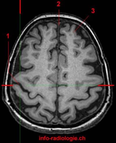
Picture 8 : MRI of the brain in T1-weighted axial view
1) Central sulcus, 2) Longitudinal cerebral fissure, 3) Superior frontal gyrus
MRI Source: w-radiology.com

Picture 9 : MRI of the brain in T1-weighted coronal view
1) Central sulcus, 2) Lateral ventricle, 3) Cerebellum
MRI Source: w-radiology.com
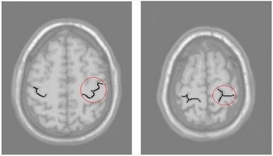
Picture 10 : The “omega” (Pleft image) and “lambda” (right image) shapes of the central sulcus.
Source: Zuo Wei, Identification and Segmentation of the Central Sulcus from Human Brain MR Images, National University of Singapore, 2004
Clinical Importance: What are the Functions of the Central Sulcus?
The central sulcus is much appreciated in the anatomical sense. It serves as the most important landmark in identifying the other parts of the cerebral cortex. It separates the frontal lobe and parietal lobe. It divides the sensory and motor areas of the brain [4].
The identification of central sulcus is extremely important in neurosurgery. It serves as a guide in determining the cortical areas of the brain. It is important in the procedure of removing brain lesions associated with the somatosensory cortex. Also, it is a relevant anatomical structure in avoiding functional deficits to patients who have to undergo epilepsy surgery.
If the neurosurgeon is not able to clearly identify the central sulcus, the patient will be at risk for motor, intellectual, memory, and personality problems. The frontal lobe may have disturbances in its functions [3].
Researchers believe that the shape of the central sulcus is somehow related to the dominant hand that we use. The dominant hand has a larger cortical area than the nondominant hand. It was found that the non-symmetrical features of central sulci of the left and right hemispheres of the brain are a lifelong process wherein age, gender, and usage or handedness are factors that contribute to its depth [5, 6].
In a study published in Oxford Journals, there is a difference in the sulcal depth in males and females. For males, there is leftward asymmetry in the superior end of the sulcus and for females, the asymmetry is observed along the midpoint of central sulcus. It was also found that neuroplasticity in males is greater than that of the females [5].
References
- Waxman SG, Clinical Neuroanatomy 26th edition, McGraw-Hill 2010, p 131
- White JS, USMLE Roadmap Neuroscience 2nd edition, McGraw-Hill 2008, p 201
- Zuo Wei, Identification and Segmentation of the Central Sulcus from Human Brain MR Images, National University of Singapore, 2004
- Ono M et al, Atlas of the Cerebral Sulci, Thieme 1990, pp 36-41
- Cykowski MD et al, The Central Sulcus: an Observer-Independent Characterization of Sulcal Landmarks and Depth Asymmetry, Oxford Journals Volume 18 Issue 19
- Hugdahl K & Westerhausen R, The Two Halves of the Brain, MIT Press 2010, pp 155-157
- Central Sulcus (Fissure of Rolando) accessed on http://w-radiology.com/central-sulcus.php