What is the Mastoid Process and Function
The mastoid part of the temporal bone houses the mastoid process. Mastoid process is the bony prominence easily felt behind the earlobe.
It is one of the key features of the lateral cranium. It is located behind and below the external auditory meatus. It primarily functions as attachment to the neck muscles.
Inside of it are mastoid air cells or sinuses that are prone to inflammation and infection. These are cells are connected to the middle ear. Mastoid air cells are covered by mucoperitoneum that is continuous with the tympanic cavity and with the squamous part of the temporal bone [1].
The mastoid notch is located medial to the mastoid process. It is pierced by stylomastoid foramen in front and mastoid foramen is found on its back. Mastoid notch serves as the site of muscle attachment for the anterior and posterior bellies of the digastrics whose function is to open the mouth [2].
At birth, mastoid process may not be palpated. Its protrusion is due to the pulling of sternocleidomastoid muscle of the neck when a person starts to move his head. It only appears as a bony projection once the child turns 2 [3].
Where is the mastoid process ?
Photos of Anatomy and Location
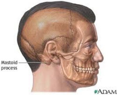
Picture 1: Site of Mastoid Process
Image Source: pennmedicine.org
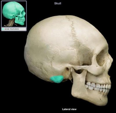
Picture 2 Lateral view of the skull highlighting the mastoid process.
Image Source: studyblue.com
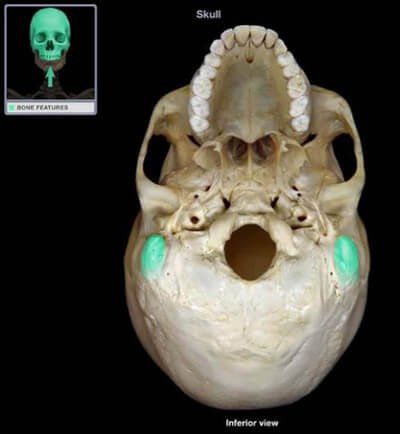
Picture 3 : Inferior view of the skull highlighting the mastoid process.
Image Source: studyblue.com
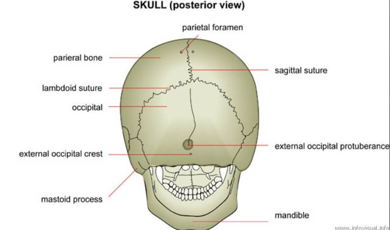
Picture 4: Posterior View of the Skull showing the Mastoid Process
Image Source: gl1800riders.com
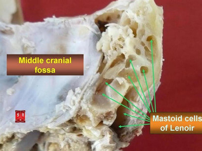
Picture 5: Mastoid Cells of Lenoir
Image Source: wikipedia.org
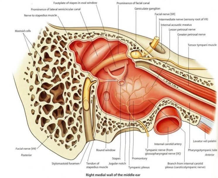
Picture 6: Mastoid Process and its Relationship with the Middle Ear
Image Source: studyblue.com
Clinical Importance
No Mastoid Process in Newborns
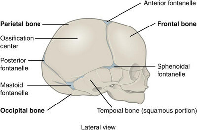
Picture 7: Lateral skull of a newborn shows no mastoid process.
Image Source: cnx.org
A newborn has no mastoid process. This means that there is less protection for the facial nerve or CN VII after birth. This nerve arises from the stylomastoid foramina and since there is no mastoid process yet, it develops close to the surface. This makes it prone to injury or damage during surgical procedures such as forceps delivery or operations for treating middle ear problems.
The mastoid process starts to develop after the patient turns 1 year old. That is when the sternocleidomastoid muscles pull on the petromastoid parts of the temporal bones. Mastoid process is already developed by the age of 2 [4].
How Infection Spreads from the Middle Ear to the Brain
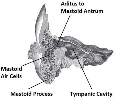
Picture 8 Mastoid Air Cells and the Nearby Structures
Image Source: teachmeanatomy.info

Picture 9: Note the close proximity of the abscessed mastoid antrum to the sigmoid sinus and cerebellum.
Image Source: smokh.org
The mastoid antrum is a small cavity found at the back of the petrous part of temporal bone. By way of aditus, it serves as a bridge between the mastoid air cells, posterior wall of the middle ear, and the sigmoid sinus and cerebellum of the brain. It is through the mastoid antrum that infection is spread from the ears to the brain [1].
Mastoiditis
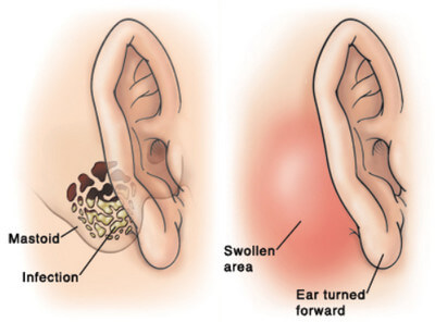
Picture 10: Mastoiditis is caused by bacterial infection causing the ear, particularly the posterior area, to appear swollen and enlarged.
Image Source: fairview.org
Mastoiditis is the inflammation and infection of the mastoid bone. This disease is common among children and was once known as one of the most common cause of mortality among them primarily because medications find it hard to reach its target, which is the mastoid bone. Furthermore, the infection could spread to the brain.
Mastoiditis is caused by ear infections, particularly by otitis media. As explained earlier, the mastoid air cells are connected to the middle ear. It can also be caused by cholesteatoma, which will be discussed further in the next sections of this article.
Signs and symptoms of mastoiditis include ear pain, ear drainage, swelling and redness around the ear, enlarged ear, impaired hearing, fever, and headache.
Treatments for mastoiditis include antibiotics, ear drops, and ear cleaning. Myringotomy (fluid drainage of the middle ear) and mastoidectomy (removal of the infected mastoid bone) are options if the disease is severe.
Mastoiditis should not be left untreated because it in the long run, it might result to hearing loss, meningitis, or brain abscess that can lead to death [5, 6].
Parotiditis and Mumps
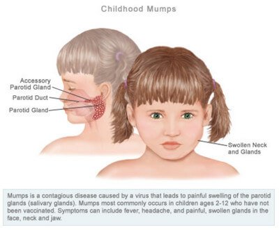
Picture 11: Note the proximity of the infected site to the mastoid process.
Image Source: healthimpactnews.com
Inflammation and infection of the parotid gland causes severe pain that is aggravated when chewing. The parotid sheath that surrounds the parotid gland is tough and when the gland swells, the sheath limits its swelling, thereby producing severe pain.
The mastoid process comes into the picture when the pain is aggravated when opening the mouth and chewing. As the mouth opens, the posterior border of the mandibular ramus moves downwards and backwards, compressing the mastoid process. This is the reason why it hurts to eat when you have parotiditis or mumps [4].
Cholesteatoma
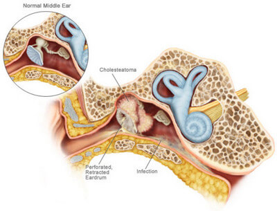
Picture 12: Cholesteatoma
Image Source: chroniclescamera.blogspot.com
Cholesteatoma is an ear disease wherein a benign skin cyst pervades the mastoid process and the middle ear. This occurs when the Eustachian tube, which normally equalizes the ear pressures, does not open enough to perform its function. As a result, the tympanic membrane or eardrum becomes perforated and retracted as it forms a pocket that accepts the skin cyst.
The patient with cholesteatoma has a history of chronic ear infections. Alongside with this, there is ear discharge and gradual hearing loss.
Cholesteatoma may be treated through surgical procedures such as tympanoplasty, mastoidectomy, or ossiculoplasty [6].
Mastoid Cancer
Malignant lumps and tumors of the mastoid are mostly due to squamous cell carcinoma. It is a form of skin cancer. People who have chronic mastoid infection, family history of skin cancer, and exposure to coals, tars and arsenics are more prone to develop mastoid cancer [6].
References:
- Ellis H, Clinical Anatomy 11th edition, Blackwell Publishing Ltd., 2006, p 386
- Saladin K, Anatomy & Physiology: The Unity of Form and Function 5th edition, The McGraw-Hill Companies Inc., 2009, p 252
- Snell RS, Clinical Anatomy by Regions 9th edition, Lippincott Williams & Wilkins, 2012, p 663
- Moore KL et al, Clinically Oriented Anatomy 6th edition, Lippincott Williams & Wilkins, 2010, pp 840 926
- http://www.webmd.com/cold-and-flu/ear-infection/mastoiditis-symptoms-causes-treatments
- http://www.livestrong.com/article/190151-diseases-of-the-mastoid/

