The Heart
Heart is a very important organ which acts as a muscular pump.
It pumps the blood that circulate around the whole body and provide oxygen and nutrients to all organs which is needed for their survival. The heart is a conical organ and the size of the heart depends on the age of the person which is roughly the size of a closed fist of each person.
The heart beats rhythmically pumping the blood right from birth to death of a person and each pumping action causes a rhythmic contraction and expansion of the heart. [1,2]

Picture 1: Anterior view of chest wall
Image Source: inkling.com
Location of the Heart
Location of the Heart in Thorax
Heart is located safely inside the chest cavity which looks like a cage bound by the ribs and breast bone (sternum). The chest cavity is also called as thoracic cavity and it lies between neck and abdomen. Heart lies slightly to the left in the chest cavity. [2]
Boundaries of thorax:
- At the back –12 thoracic vertebra forming the part of back bone
- At the front – breast bone or sternum
- Enclosed by ribs which starts from the vertebral column at the back to the sternum in the front on both sides and the muscles between the ribs (intercostal muscles)
- Above – thoracic inlet – the opening at root of neck in which blood vessels and nerves enter and leave the chest cavity [6]
- Below – diaphragm – the muscular organ that separates the chest cavity from abdomen [5].
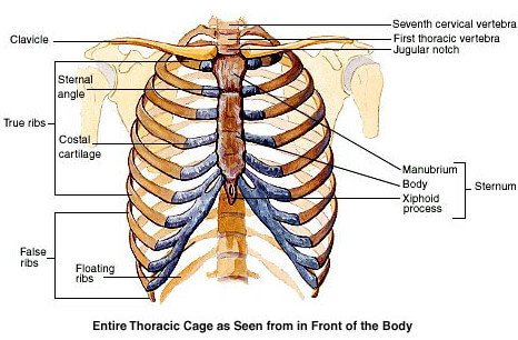
Picture 2: Thoracic cage
Image Source: mhhe.com
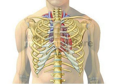
Picture 3: Location of heart inside chest cavity
Image Source: dccdn.de
Location of heart in the Mediastinum
Heart is located in a space in the centre of the chest cavity called mediastinum in particular theMiddle mediastinum. It is the space between the 2 lungs and the heart is wedged between them. [3]
The mediastinum is divided into
- Superior mediastinum
- Inferior mediastinum.
Inferior mediastinum is further divided into
- Anterior mediastinum
- Middle mediastinum
- Posterior mediastinum

Picture 4: Division of mediastinum
Image Source: medicine.academic.ru
Boundaries of the Mediastinum
- In the front – sternum
- At the back – vertebral column
- Above – Thoracic outlet – the opening of the chest cavity at the top
- Below – Diaphragm – the muscular layer separating the chest from the abdomen
Boundaries of Middle Mediastinum:
- In the front – Inner surface of the sternum
- At the back – esophagus which is the muscular tube that carries the food to the stomach
- On both sides –the pleura – layer of tissue lining the lungs
- Above – Superior mediastinum
- Below – diaphragm
Along with the heart enclosed in its pericardium, the great vessels like aorta, pulmonary trunk, Superior venacava, arch of azygos vein and main bronchi are also present in the middle mediastinum.

Picture 5: Heart in the middle mediastinum
Image Source: fastbleep.com
Location of heart in Pericardium
During each pumping action the heart expands and contracts forming a pulsation. Heart lies in a sac like covering called pericardium which makes pulsations smoother without any strain [4]. This pericardium consists of 2 layers with a lubricating fluid in between called the pericardial fluid. The outer layer is called as fibrous pericardium. It has strong attachment with the diaphragm below and it merges with the walls of great blood vessels like aorta and pulmonary arteries that arise from the heart and pulmonary veins, superior and inferior venacava that reach the hear. It restrains the heart from overexpanding. The inner layer is called as serous pericardium which inturn is made up f 2 layers and covers the heart like a blanket. [7]
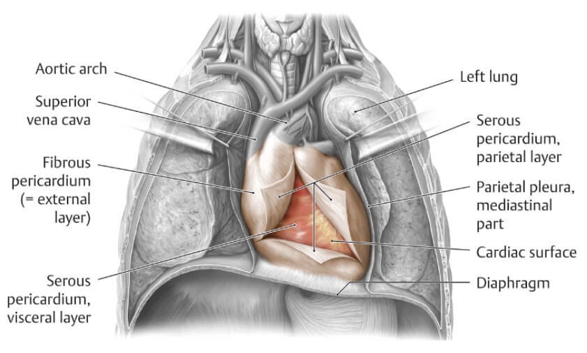
Picture 5: Heart inside the pericardium
Image Source: inkling.com
Surface Anatomy of Heart
The border of the heart that lies inside can be traced on the front of chest wall by marking the following points:
1. Mark a point 1cm from the right sterna line along the Upper border of 3 rd right costal cartilage.
2. Mark another point at 2.5 cm from the left lateral sterna line along the lower border of the second left costal cartilage Joining points 1 and 2 forms superior border of heart.
3. A point 2 cm to the right of the sternum along the space between 6th and 7th rib on the right side where the cartilage joins the sternum.
4. Mark a point 9 cm to the left of midsternal line. This point lies corresponding to the apex of heart inside Joining points 3 and 4 forms the inferior border of heart
Thus right border is formed by joining 1 and 3 and left border of heart is formed by joining 2 and 4
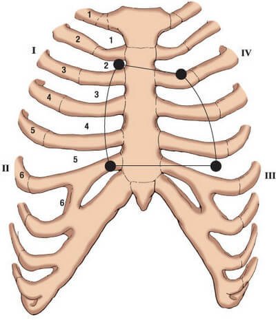
Picture 6: Surface anatomy of heart
Image Source: web.uni-plovdiv.bg
Location of Heart in relation to other organs
Anterior relations of heart from front to back
- Skin
- Connective tissues
- Anterior chest muscles
- The ribs 2 to 7 on left side
- Sternum
- Structures of anterior mediastinum
Posterior relations of heart
- Esophagus
- Thoracic duct
- Descending aorta
- Azygos vein
- Other structures of posterior mediastinum
On either side, heart is wedged between the lungs and the structures that are present in the medial side of the lungs : [7]

Picture 7: Posterior relations of heart
Image Source: cloudfront.net

Picture 8: Transverse section at the level of heart showing its relation with other organs
Image Source: www.biosbcc.net
Location of Heart – Exceptions
Dextrocardia
We all know that heart is located in the left side of the chest. But in some rare cases, the heart rotates and the apex points towards the right side of chest. This happens due to a change that happens during the development in which the growing heart flips to the other side and continues growing and the cause is unknown. This condition is known as dextrocardia. It is mostly associated with mirror images of the organs in the digestive system and genitourinary system. The person will have other abnormalities associated with heart problems and is confirmed by taking a plain chest radiograph. Some people lead a normal life without serious problems while it may be life threatening when other organs are involved. [9, 10]

Picture 9: Radiograph showing dextrocardia
Radiograph Source: media.tumblr.com
Enlarged heart
There are some medical conditions that cause the enlargement of heart. Mostly the enlarged heart is associated with heart failure [12]. The enlargement causes change in the location of heart by extending its borders to more outward, downward direction and occupies more space on the right side of chest as well. The heart appears like a big balloon. These conditions include high blood pressure, heart valve diseases, hypertrophic cardiomyopaty, cardiomegaly, pericardial effusion etc [11]
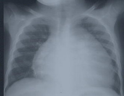
Picture 10: Radiograph Showing Cardiomegaly – the Heart literally occupies the entire middle mediastinum
X-Ray Source: bipai.org
References:
- http://www.webmd.com/heart/picture-of-the-heart
- http://en.wikipedia.org/wiki/Heart
- http://www.texasheartinstitute.org/HIC/Anatomy/anatomy2.cfm
- http://anatomy.uams.edu/anatomyhtml/viscera_thorax.html
- Cunningham’s manual of practical anatomy 15/e vol2 pg 1
- https://www.inkling.com/read/essential-clinical-anatomy-keith-moore-4th/chapter-1/thoracic-wall
- http://www.dartmouth.edu/~humananatomy/part_4/chapter_23.html
- http://www.auhsd.us/view/8309.pdf
- http://www.nlm.nih.gov/medlineplus/ency/article/007326.htm
- http://www.cardiacmatters.co.uk/heart-condition-dextrocardia-explained.html
- http://www.mayoclinic.org/diseases-conditions/enlarged-heart/basics/causes/con-20034346
- http://www.heart.org/HEARTORG/Conditions/HeartFailure/AboutHeartFailure/Enlarged-Heart_UCM_450777_Article.jsp