
Cricoid Cartilage Definition and Structure
Cricoid cartilage is an unpaired hyaline cartilage located in the larynx at C6 vertebral level. It rests inferior to the thyroid cartilage.
It has a characteristic signet ring shape where the anterior part has an arch and the posterior part is a broad lamina. Cricoid cartilage increases its size as it goes posteriorly. Anterior size is the narrowest measuring 0.5 cm. Laterally, it increases to 1 cm. The lamina is the broadest measuring about 2.5 cm [1].
The inferior cornu articulates at the lateral sides of the cricoid cartilages via facets. On the posterior portion, it articulates with the arytenoid cartilages. All articulations with the cricoid cartilage are synovial [2].
Cricoid cartilage is attached to the thyroid cartilage in the presence of cricothyroid ligament. There is also a cricotracheal ligament that attaches the cricoid cartilage to the first ring of the trachea [3].
Anteriorly, cricoid cartilage gives rise to the straight and oblique heads of cricothyroid muscle. On its superior surface near the arytenoid cartilages, it gives rise to the lateral and posterior cricoarytenoid muscles [1].
Cricoid cartilage is one of the six laryngeal cartilage types. The other five are the following:
- Epiglottis: This is attached to the thyroid gland. It is made up of fibrocartilage with a shape like that of a spoon. Epiglottis and cricoid cartilage are the laryngeal cartilages that are not paired.
- Thyroid: This is commonly known as the Adam’s apple. It consists of two hyaline laminae.
- Arytenoid: This is a triangular paired cartilage that surrounds the cricoid cartilage.
- Corniculate: This is another paired cartilage that sits on top of the arytenoid cartilage.
- Cuneiform: This is the type that has no articulations. It is a paired cartilage that can be found on aryepiglottic folds [4].
Images of Anatomy: Structure and Location
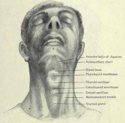
Picture 1: Location of the Cricoid Cartilage
Image Source: primehealthchannel.com
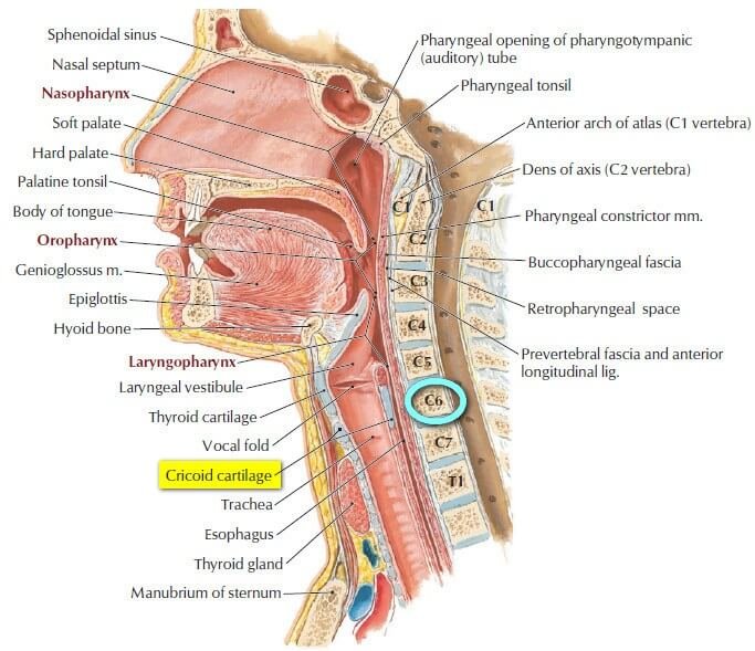
Picture 2: Cricoid cartilage at C6 vertebral level
Image Source: Hansen JT, Netter’s Clinical Anatomy 2nd edition, Saunders 2010, p 424
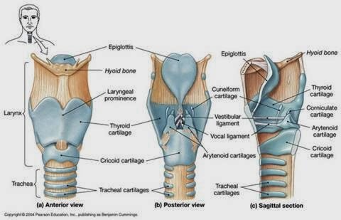
Picture 3: The cricoid cartilage in anterior, posterior, and sagittal views.
Image Source: smallcollation.blogspot.com
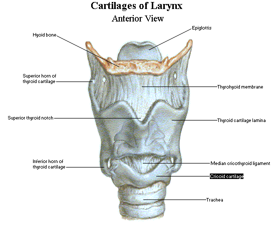
Picture 4: Laryngeal Cartilages
Image Source: accweb.itr.maryville.edu
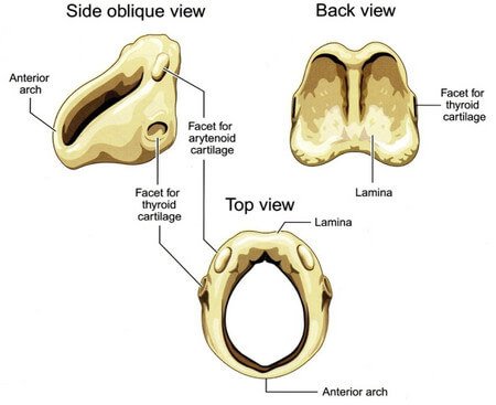
Picture 5: Different views of the cricoid cartilage.
Image Source: studyblue.com
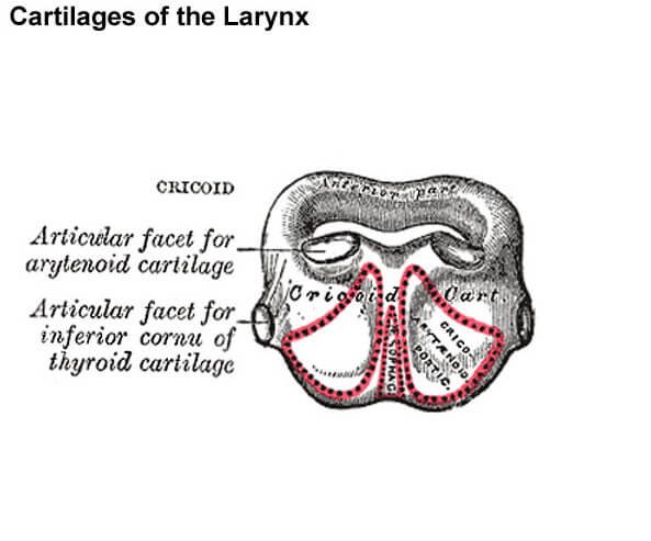
Picture 6: Facets of Cricoid Cartilage
The superior facets articulate with the arytenoid cartilages. The inferolateral facets articulate with the inferior cornu of thyroid cartilage.
Image Source: embryology.med.unsw.edu.au
Cricoid Cartilage Functions
The main function of the cricoid cartilage is to anchor the muscles, ligaments, and cartilages that are attached to it in order to facilitate the opening and closing of epiglottis. It has muscle fibers attached to it that allow the food bolus to pass from the pharynx to the esophagus. It also helps in production of speech or sounds [5, 6].
Clinical Significance
Cricothyrotomy
In the event that airway is not established by all other means, emergency cricothyrotomy may be done to gain access through the trachea and provide respiration.
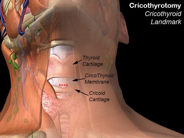
Picture 7: The thyroid and cricoid cartilages serve as a landmark to identify the cricothyroid membrane which will be incised during cricothyrotomy.
photo Source: fpnotebook.com
Look for the cricothyroid membrane by determining the location of thyroid notch and the cricoid cartilage. Cricothyroid membrane is in between them, as depicted in Picture 7.
Once identified, cricothyroid membrane is incised and a tube must be placed immediately for it to remain open. If done properly, you have saved a person’s life by establishing airway.
There are cases when a patient has pyramidal lobe that stems off the thyroid gland. This occludes the area for cricothyrotomy. When the pyramidal lobe is being cut, the patient will be at risk for haemorrhage [4].
Sellick’s Maneuver or Cricoid Pressure
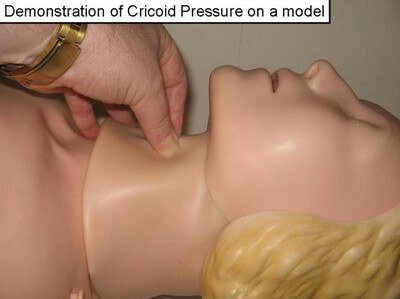
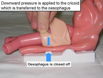
Picture 9: Sellick Maneuver or Cricoid Pressure
Image Source: facs.med.cuhk.edu.hk
Sellick’s maneuver or cricoid pressure is performed during endotracheal intubation or rapid sequence induction (RSI) to prevent aspiration, thereby ensuring entry of oxygen into the lungs and not the stomach.
Behind the cricoid cartilage is the esophagus. The thumb and the index finger are used to apply pressure on the cricoid cartilage in order to close off the esophagus.
However, studies have found that Sellick’s maneuver poses risks to the patient and problems to the practitioner.
- Proper visualization of the trachea is obscured when applying pressure to the cricoid cartilage.
- In some cases, airway obstruction was worsened when cricoid pressure was performed.
- Cricoid cartilage fracture may happen when too much pressure is applied.
It was argued that Brian Sellick had a small sample and limited evidence when he proved that cricoid pressure is needed in performing endotracheal intubation and RSI. Well, its use is entirely up to the case. If Sellick’s maneuver helps both the patient and practitioner, do it. If it worsens the problem, then don’t [7].
Fracture of Laryngeal Cartilages:
The most commonly fractured laryngeal cartilages are thyroid and cricoid cartilages. Physical injury or trauma is usually the reason for having fracture on these parts. The patient develops laryngeal inflammation, swelling, or even hemorrhage. Possible complications are problems in speech and airway obstruction [1].
References:
- Clemente CD, Clemente’s Anatomy Dissector, Lippincott Williams & Wilkins 2010, p 416-424
- Snell RS, Clinical Anatomy by Regions 9th edition, Lippincott Williams & Wilkins 2012, p 645
- Tortora GJ & Derrickson B, Principles of Anatomy & Physiology 13th edition, John Wiley & Sons Inc. 2012, p 923
- Hansen JT, Netter’s Clinical Anatomy 2nd edition, Saunders 2010, p 424-427
- http://www.healthline.com/human-body-maps/cricoid-cartilage
- Kent RD, The MIT Encyclopedia of Communication Disorders, MIT Press 2004, p 14
- Bledsoe BE, Sellick’s Maneuver — Not the Panacea We Thought accessed on http://www.ems1.com/ems-products/education/articles/333130-Sellick-s-Maneuver-Not-the-Panacea-We-Thought/