Sternum Definition and Function
The sternum or breastbone is a vertical flat bone lying at the anterior middle part of the chest.
Externally, it is made up of compact bone. Inside of it is a cancellous or spongy tissue that is entirely vascular.
The adult length of the sternum measures 15-17 cm. It is larger in males than in females. Sternum protects the organs inside the mediastinum, especially the heart [1, 2, 3].
Parts of the Sternum
Manubrium
- The manubrium sterni is the most superior part of the sternum that lies at the levels of 3rd and 4th thoracic vertebrae.
- The superior border of the manubrium has the broadest structure among the other parts of the sternum.
- On its center is the suprasternal or presternal or jugular notch. This is the depressed part that can be palpated in between the clavicles.
- The lateral sides of the superior border of the sternum articulate with the clavicles via sternoclavicular joints.
- The lateral borders have facets that articulate with the ribs.
- The inferior border has a cartilage that articulates with the sternal body at manubriosternal joint.
- The anterior surface serves as an attachment to the pectoralis major and sternocleidomastoid muscles.
- The posterior surface serves as an attachment to the sternohyoid and sternothyroid muscles.
Body
- The corpus sterni or gladiolus is the longest part of the sternum, although it is narrower than the manubrium. The body of the sternum is found at the level of T5-T9 vertebrae.
- The anterior surface of the sternal body is almost flat. It is where the pectoralis major is attached.
- There are three horizontal ridges that pass through the 3rd, 4th, and 5th articular facets. Between the 3rd and 4th parts of the sternal body, the sternal foramen can be seen.
- The posterior surface also has these three lines. On its lower end is the origin of the transversus thoracis.
- The superior border of the sternum articulates with the manubrium through the manubriosternal joint.
- The gap between the two is called the sternal angle or angulus Ludovici or angle of Louis, named after Antoine Louis.
- The lateral border has articular depressions on which the 2nd to 7th costal cartilages are attached.
- The inferior border is the narrowest part of the manubrium and it articulates with the xiphoid process through the xiphisternal joint which lies at the level of the 9th thoracic vertebra.
At birth until young adulthood, there are four sternebrae attached to sternal synchondroses. When a person reaches 25 years, these sternebrae fuse to form three synostoses.
Xiphoid Process
The xiphoid process, also known as processus xiphoideus, ensiform or xiphoid appendix, is the smallest part of the sternum. It lies at the level of T10 vertebra. Costal cartilages nor ribs are attached to it.
It is made up of cartilage until it ossifies at the age of 40 and in some cases, they become one with the sternal body in old age.
- The superior border articulates with the sternal body via xiphisternal joint.
- On its sides are articular facets that provide attachment to the 7th rib.
- The inferior border gives rise to the linea alba.
- The anterior surface is where the anterior costoxiphoid ligament and a part of rectus abdominis are attached.
- The posterior costoxiphoid ligament, transversus thoracis, and diaphragm are attached to the posterior surface.
- The lateral borders are connected with abdominal aponeuroses [1, 2, 4].
For more information : please check – Xiphoid Process Pain
Anatomy Images: Where is the Sternum Located?

Picture 1: Sternum is a part of the skeletal system. It lies on the anterior thoracic wall in the middle. Its parts are the manubrium (green), body (blue), and xiphoid process (violet).
Image Source: wikipedia.org

Picture 2: Sternum, Costal Cartilages, and Ribs
Image Source: newhealthguide.org

Picture 3: Anterior View of the Thoracic Cage
Image Source: antranik.org

Picture 4: Anterior and Lateral Views of the Sternum
Image Source: paradoja7.com
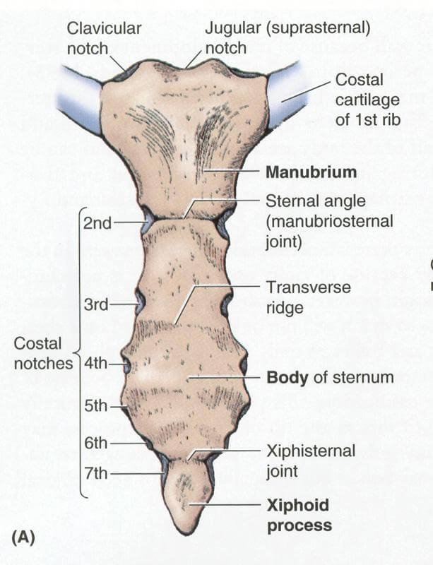
Picture 5: Anterior View of the Sternum
Image Source: oxford174.com
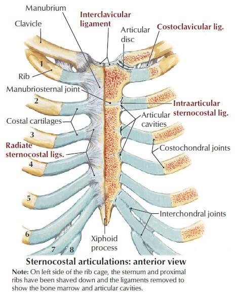
Picture 6: Sternocostal Articulations
Image Source: Clinical Anatomy 2nd edition
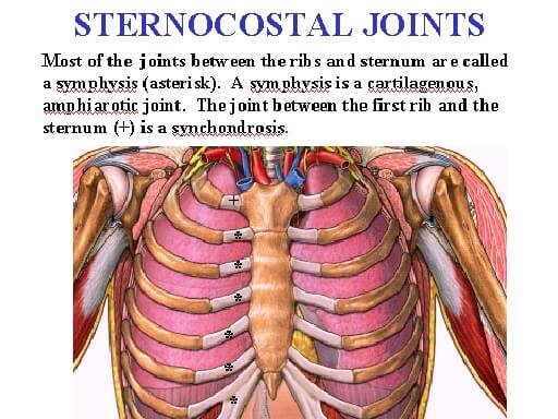
Picture 7: The first sternocostal joint is categorized as synchondrosis. The succeeding joints are symphysis.
Image Source: faculty.ucc.edu
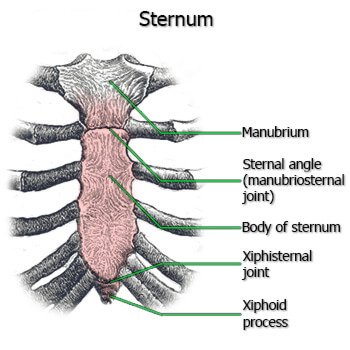
Picture 8: Joints of the Sternum
Image Source: healthguidance.org
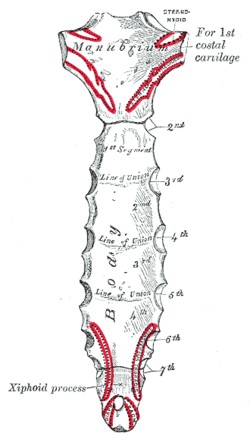
Picture 9: Posterior Surface of the Sternum and Articulations with the Costal Cartilages
Image Source: wikipedia.org
Clinical Importance of Sternum
Sternal Angle or Angle of Louis as a Landmark
Sternal angle or the angle of Louis is the articulation that is formed by the fusion of manubrium and body of the sternum. This is a very important landmark since it is where the costal cartilages and ribs are first counted. It rests at the level of 2nd costal cartilage and in between 4th and 5th thoracic vertebrae.
Xiphoid Process as a Landmark
Xiphoid process may be small but it is an important landmark for the structures beneath it. It serves as a landmark for:
- The anatomical end for anterior thoracic cage
- Formation of infrasternal or subcostal angle together with the subcostal margins
- Inferior border of the heart
- Superior border of the liver
- Diaphragm’s central tendon
Bone Marrow Biopsy
The sternum is a hematopoietic tissue, meaning blood cells are produced in this site. To obtain a bone marrow biopsy, a large bore needle is introduced into the sternum and a sample is aspirated [4].
Surgery
A bone saw is used to gain access to the thoracic cavity in surgeries involving the heart, great vessels, thymus gland, and retrosternal goiter. Wire sutures are used to close the sternum in surgery.
Sternal Fracture
The costal cartilages cushion the sternum and protect it from injury or trauma. However, too much force cannot be handled by the sternum and that is where fracture and dislocation set in.
If the rescuer’s hands are placed wrongly on the chest of the patient during cardiopulmonary resuscitation (CPR), the xiphoid process might be fractured and pierce the underlying organs.
There may be a sensation of a “popping” sternum in sternal fracture. The manubrium may be displaced from the sternal body. Fracture of the sternum may also be associated with fracture of the thoracic vertebrae [3, 5].
Sternum Pain
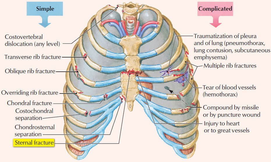
Picture 10: Thoracic Cage Injuries
Sternal fracture is a form of a simple thoracic cage injury. Although simple, it should still be given immediate medical attention.
Image Source: Netter’s Clinical Anatomy 2nd edition, Saunders, 2010
Causes of Sternum pain
Fractures, dislocations, joint separations, wounds, or any other forms of injury or trauma to the thoracic cage cause pain to the sternum.
Sternum is located in the middle of the chest cavity. The surrounding anatomical structures are attached to it. So if these become damaged, no matter how simple the diagnosis may be, there will be a resulting sternal pain.
Respiration is an involuntary, essential function. It involves spontaneous, continuous movement of the thoracic cage, including the sternum.
If the sternum is broken, common sense will tell that pain results. There will be pain in your sternum with each breath you take.
Also see : Causes of Pain under right rib cage
Treatment for Sternum pain
To deal with sternum pain, nerve block is performed wherein the intercostal nerve that transmits pain signals to the brain is being anesthetized [6].
References:
- Gray H, Anatomy of the Human Body, Bartleby Bookstore
- Moore KL et al, Clinically Oriented Anatomy 6th edition, Lippincott Williams & Wilkins 2010, pp 76-78
- Tortora GJ & Derrickson B, Principles of Anatomy & Physiology 13th edition, Biological Science Textbooks Inc. 2012, p 245
- Snell RS, Clinical Anatomy by Regions 9th edition, Lippincott Williams & Wilkins 2012, p 35
- Ellis H, Clinical Anatomy 11th edition, Blackwell Publishing 2006, p 11
- Hansen JT, Netter’s Clinical Anatomy 2nd edition, Saunders 2010, p 76
