
Endosteum : Definition and Functions
The endosteum is a structure in the middle of bone tissue and bone marrow. It is a thin covering that surrounds the medullary cavity.
It coats the inner compact bone and the trabeculae of the spongy bone. It covers the loose structures found inside the bone. It is made up of connective tissue and one layer of cells.
The osteogenic capability of the endosteum lies on the fact that it houses osteoprogenitor cells or preosteoblasts that differentiate into osteoblasts (bone-forming cells), bone matrix-secreting cells, or bone-lining cells.
Osteoprogenitor cells are often called endosteal cells. It is hard to recognize even under the microscope. It is described to be flat with elongated nucleus. The features of the cytoplasm are vague.
Together with the periosteum, endosteum is responsible for growth of the bone in diameter but contrary to the periosteum, the fibrous layer of the endosteum is indistinct [1–5].
Photos of Anatomy and Histology

Picture 1: Periosteum vs. Endosteum
Image Source: wikipedia.org
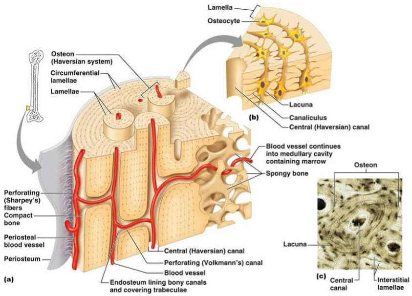
Picture 2: Skeletal tissues showing how endosteum lines the body canals and covers trabeculae
Image Source: classes.midlandstech.edu

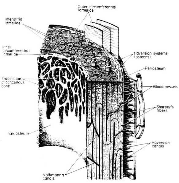
Picture 3: Parts of the Skeletal Tissue
Image Source: rci.rutgers.edu
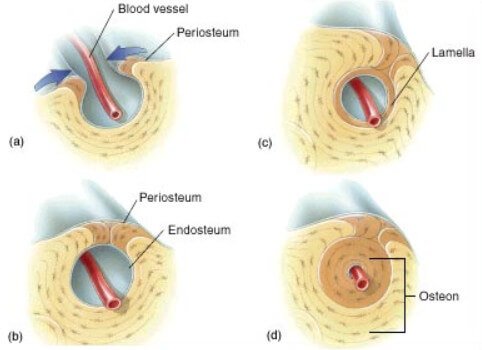
Picture 4: Endosteum and Other Parts of the Bone
Image Source: knowyourbody.net
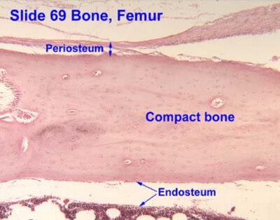
Picture 5: Endosteum on Cross-Section
Image Source: mhhe.com
Picture 6: Histology of the bone showing periosteum and endosteum
Image Source: bvetmed1.blogspot.com
Clinical Importance
Chemical exchange between the bone marrow and blood vessels becomes possible through the osteoblasts of the endosteum.
Endosteal cells are active in the process of bone growth, repair, and remodelling. There are parts of the bone matrix that are not entirely covered by the endosteum. These exposed areas are the attachment sites for osteoblasts and osteoclasts wherein they can deposit or remove components of the bone matrix.
Endosteum and periosteum contribute to bone repair and reconstruction after a fracture occurs. Blood vessels and tissue surrounding the injured area bleed and eventually form a clot through the edges of the broken bone.
The cells of endosteum and periosteum undergo rapid mitosis and proliferation then they migrate towards the affected site. There will be formation of a new bone that will serve as a bridge which temporarily stabilizes the broken ends of the bone [6, 7].
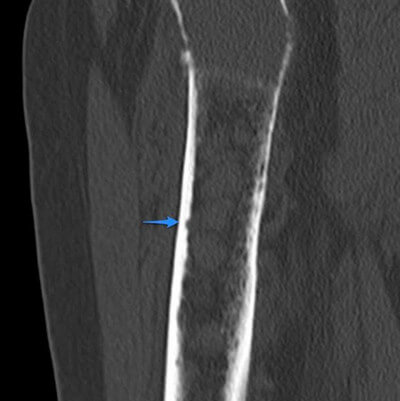
Picture 7: Endosteal Scalloping
Image Source: radiopaedia.org
If medullary lesions develop along the inner aspect of the cortical bones, especially in the long bones, endosteal scalloping may be observed. Although these medullary lesions may be slow-growing and non-infiltrating, endosteal scalloping is associated with both benign and malignant conditions which include enchondroma, osteomyelitis, chondromyxoid fibroma, skeletal amyloidosis, periprosthetic osteolysis, brown tumor, chondrosarcoma, multiple myeloma, and skeletal metastasis [8].
In nuclear medicine radiation dosimetry, a normal endosteum measures 10 µm in thickness. Endosteal thickness of 50 µm from the medullary cavity and trabeculae suggest sensitivity of radioactive cells which makes a person prone to bone cancer [9].
Endosteum vs. Periosteum
Periosteum and endosteum are both parts of the bone. Aside from their rhyming words, many are still puzzled with the difference of these two. Here is a table summarizing these two very important parts of the bone [10].
Table 1: Comparison between Endosteum and Periosteum
References
- Ross MH & Pawlina W, Histology: A Text and Atlas 6th edition, Lippincott Williams & Wilkins, 2011, p 221
- Tortora GJ & Derrickson B, Principles of Anatomy and Physiology 13th edition, Biological Science Textbooks Inc., 2012, p 184
- Johnson KE, Histology and Cell Biology 2nd edition, Williams & Wilkins, 1991, pp 112-114
- Eroschenko VP, diFiore’s Atlas of Histology with Functional Correlations 11th edition, Lippincott Williams & Wilkins, 2008, p 90
- Henrikson RC & Mazurkiewicz JE, Histology Volume 518, Lippincott Williams & Wilkins, 1997, p 131
- Khan R, A Textbook of Biotechnoloy Volume 2, Firewall Media, 2007, pp 62-65
- Martini F et al, Anatomy and Physiology, Rex Bookstore Inc., 2007, pp 135 & 144
- http://www.ehealthstar.com/anatomy/endosteum
- McParland BJ, Nuclear Medicine Radiation Dosimetry, Springer Science and Business Media, 2010, p 512
- Weerakkody Y & Gaillard F, Endosteal Scalloping accessed on http://radiopaedia.org/articles/endosteal-scalloping