Introduction
For centuries, cadaver dissection has been a cornerstone of medical education, providing hands-on anatomical knowledge
. Yet cadavers are expensive, limited in supply, and can’t simulate living physiology. Enter holographic patients—advanced 3D projections that allow medical students and professionals to visualize and manipulate virtual anatomy in real time.
Driven by developments in augmented reality (AR) and holographic imaging, this new technology promises to supplement or even replace traditional cadaver labs for certain training exercises.
How close are we to seeing these digital figures in every medical classroom? This article dives into the cutting-edge of holographic medical training, its potential benefits, and the hurdles to mainstream adoption.
Why Holographic Patients?
Limits of Cadaver Dissection
- Ethical and Cost Concerns: Many institutions face scarcity or high costs for sourcing and maintaining cadavers.
- Lack of Physiological Dynamics: Cadavers can’t replicate blood flow, muscle contraction, or living tissue properties.
- Student Engagement: Dissection can be messy, potentially off-putting, and not all learners respond well to physical lab experiences.
The Holographic Advantage
Holographic systems promise:
- 3D Interaction: Students can rotate, zoom in, or highlight structures from any angle.
- Dynamic Physiology: Some simulations depict a beating heart, blood flow, or real-time respiratory movements.
- Customizable Pathologies: A single “holographic body” can be switched to show different diseases, organ variations, or surgical scenarios, which would be impossible with a single cadaver.
The Technology Behind Medical Holograms
AR/VR Platforms
Many “holographic” experiences rely on augmented reality headsets (e.g., Microsoft HoloLens). These headsets overlay 3D anatomical models onto real space,
so students see a patient’s body parts floating in front of them. Some systems use VR goggles with external motion sensors for a fully immersive environment.
Holographic Displays
True 3D volumetric displays, though rarer, project images that can be viewed from multiple angles without headsets. This approach might use light-field or laser plasma technology to create free-floating visuals. It’s still emerging but can offer a shared group experience in the lab.
Anatomy Data and Software
The foundation is high-resolution anatomical imaging (MRI, CT scans, or photogrammetry) plus advanced 3D modeling. Software then segments organs,
layers tissues, and generates realistic textures—sometimes even simulating pathologies or injuries. Coupled with interactive tools, students can dissect or annotate these digital bodies.
Advantages in Medical Education
Enhanced Learning Experience
Interactive 3D visuals can deepen spatial understanding of complex structures (e.g., the inner ear or pelvic region). Students can rehearse different angles or enlarge tiny structures, boosting memory retention and bridging knowledge gaps more effectively than 2D textbooks or static dissection.
Repeatable and Customizable
Unlike a single cadaver, which is finite and can degrade over time, a holographic model is reusable and can be “reset.”
Instructors can toggle between healthy and diseased states—like switching from normal lung tissue to advanced emphysema—in seconds, offering diverse case studies on demand.
Ethical and Resource-Efficient
Cutting reliance on real bodies helps address supply and ethical concerns about cadaver use. Additionally, it reduces the environmental and logistical burden of maintaining a cadaver lab, from preservation chemicals to disposal.
Early Implementations and Research
Universities Testing AR Labs
Select medical schools have started pilot programs using AR headsets for anatomy labs
. Surveys often report improved student engagement and comparable exam performance to traditional methods. In some cases, students find it easier to visualize 3D relationships in digital form.
Partnering with Hospitals and Tech Firms
Medical device giants and tech companies collaborate to refine holographic “patient” modules for advanced surgical planning. For example, surgeons might rehearse a complex operation on a patient’s 3D-scanned hologram, anticipating tricky angles or potential complications.
Challenges and Limitations
Hardware Costs and Accessibility
Top-tier AR headsets or volumetric displays remain expensive. Many institutions can’t fully replace cadaver labs overnight. Widespread adoption may require more affordable solutions or robust external funding.
Loss of Tactile Feedback
Cadaver-based learning fosters an understanding of tissue texture and resistance—holograms can’t replicate physical sensation. Some solutions incorporate haptic gloves or styluses, but these are less advanced and add complexity.
Realism Gaps
Current visuals may lack certain micro-details or lifelike organ “feel.” Also, replicating how tissues bleed or fluid flows in real dissection is tough. Over time, improved rendering and physics engines might partially address these gaps, but bridging reality remains a challenge.
Future Directions
Integration with Simulation
Holographic patients might link to manikin simulators. For instance, a physical mannequin can track vital signs, while an overlay shows internal processes. This synergy would combine tangible manipulation with visual internal representations.
Remote Learning and Telepresence
Medical students or instructors in different locations could share the same virtual “patient,” practicing group dissection or collaborative diagnosis. The synergy of 5G or high-speed networks can enable smooth multi-user experiences.
Potential Beyond Education
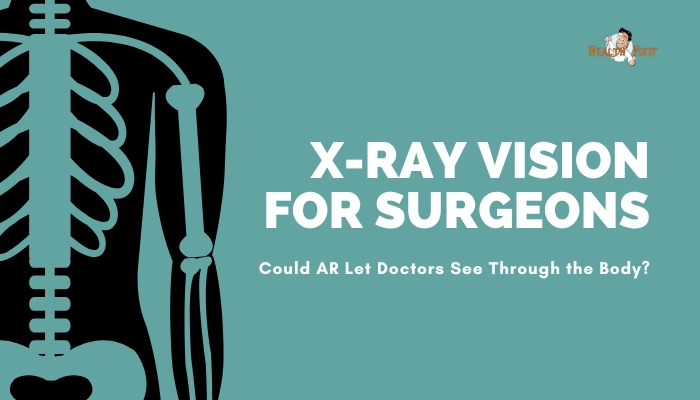
Holographic imaging might also guide real surgeries, letting a doctor see a patient’s organ maps overlayed on the operative field. This “x-ray vision” approach is emerging in AR-based surgical solutions, bridging from educational to clinical usage.
Practical Tips for Schools and Learners
- Pilot Small: If your institution is new to holographic labs, start with partial modules or single organ systems to gauge student feedback and refine hardware usage.
- Combine with Physical Training: Use holograms as a supplement, not a total replacement, especially for tactile or complex surgical skill development.
- Seek Funding and Partnerships: Collaborative efforts with edtech or healthcare companies can reduce device costs and accelerate integration.
- Assess Learning Outcomes: Compare exam scores, feedback, and skill competencies between students using holograms vs. traditional cadaver labs to refine curriculum design.
Conclusion
Holographic patients represent a frontier in medical education, promising immersive 3D anatomy that’s repeatable,
customizable, and ethically simpler than traditional cadaver labs. While challenges around cost, tactile realism, and hardware remain,
ongoing innovations in AR/VR, haptics, and advanced modeling are swiftly bridging these gaps. The future might see medical students perfecting procedures on lifelike digital bodies,
complementing or partially replacing cadaver-based training. Ultimately, the synergy of real and virtual experiences could produce better-trained doctors while respecting resource constraints and ethical concerns. As these technologies mature
, it’s plausible to envision a new golden age of high-tech, flexible, and globally accessible medical training.
References
- Azer SA, Eizenberg N. Do we really need cadavers anymore to learn anatomy in undergraduate medical education? Med Teach. 2007;29(9):913–915.
- Maresky HS, et al. Virtual and augmented reality in medical education. Med Teach. 2019;41(11):1278–1282.
- Hsieh S, et al. Interactive holographic systems for medical training: a systematic review. J Med Internet Res. 2021;23(4):e25626.
- Kenwright B, et al. Real-time volume rendering of holographic anatomy for advanced medical training. Comput Graph. 2020;90:83–93.
- Estai M, Bunt S. Best teaching practices in anatomy education: a critical review. Ann Anat. 2016;208:151–157.
- McMenamin PG, Quayle MR, McHenry CR, Adams JW. The production of anatomical teaching resources using 3D printing technology. Anat Sci Educ. 2014;7(6):479–486.
- Ferrer-Torres A, et al. Holographic augmented reality in clinical education. J Am Coll Radiol. 2021;18(2):345–350.
- Kaneko T, Seo S. Recent innovations in VR/AR technology for medical education. Int J Med Robot. 2021;17(1):e2213.
- Billings-Gagliardi S, Mazor K. Ten misconceptions about cadaver dissection. Clin Anat. 2018;31(1):4–8.
- Tam MD, Sharmila B. The potential of holoanatomy in bridging the gap in healthcare education. BMC Med Educ. 2022;22(1):478.


