Orbicularis oculi Definition
Orbicularis oculi (pronunciation: or-bi-kyu-LAR-is OK-yu-lai) refers to the muscle around the eyes.
Its origin is the medial side of the orbit and the insertion is the path that circles the orbit. The nerve responsible for it is the facial nerve or cranial nerve VII [1].
Orbicularis oculi is not attached to any bones on the lateral side that is why when you close your eyelids, they are both drawn to the middle [2].
Three Parts of Orbicularis Oculi
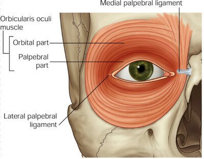
Picture 1: Orbital and Palpebral Parts of Orbicularis Oculi
Image Source: studyblue.com
Pars orbitalis or the orbital part of orbicularis oculi is the bulkiest among the three. Coarse fibers surround the entire orbit. It has two origins: the frontal bone and the maxilla. The insertion circles around the orbit. It contracts to close the eyes tight.
Pars palpebralis or the palpebral part covers the eyelid itself. It also encases the lacrimal sac and canaliculi. Compared to the pars orbitalis, it is made up of fine fibers. It originates from the medial palpebral ligament and inserts into the zygomatic bone, specifically at the lateral palpebral ligament. It acts to close the eyes gently.
Picture 2: Lacrimal Part of Orbicularis Oculi
Image Source: Stone RJ & Stone JA, Atlas of Skeletal Muscles 3rd edition, p 29
Pars lacrimalis or the lacrimal part of the orbicularis oculi is responsible for anchoring the lacrimal canal towards the eye surface. Its origin is the lacrimal bone and its insertion is the lateral palpebral raphe. It has its own ciliary bundle [2, 3].
Anatomy and Photos: Where is it Located?

Picture 3: Muscles for Facial Expression
Image Source: Tortora GJ & Derrickson B, Principles of Anatomy and Physiology 12th edition, John Wiley & Sons Inc. 2009

Picture 4: Lateral View of Facial Muscles
Image Source: Tortora GJ & Derrickson B, Principles of Anatomy and Physiology 12th edition, John Wiley & Sons Inc. 2009
Clinical Importance: What are the Functions of Orbicularis Oculi?
Orbicularis oculi is responsible for closing our eyes [1]. Examining its function is extremely important because when damage is there and it is ignored, it will eventually lead to lagophthalmos (complete failure of closing the eyes) and paralytic ectropion (eversion of the lower eyelid).
The ability of the orbicularis oculi to close the eyelids is examined by inspection. Notice the capacity of the patient to close the eyelids and determine its severity. Instruct him to close his eyes gently and tightly.
The muscle tone of the eyelids can be examined by instructing the patient to close his eyes while the practitioner counters the act. The cornea and eyelid position should also be checked [4].
Blink Reflex
When something gets into your eyes, you blink instantly. This happens because the nasociliary nerve (CN V1) senses the stimulus that will arouse the blinking reflex of both eyes. Its initiation involves the activation of motor function of the facial nerve (CN VII). This will prompt the orbicularis oculi, particularly the palpebral part, to contract, thus closing the eyes.
A part of physical examination includes the test for corneal blink reflex. The practitioner wisps cotton on your eye. If you do not blink, there might be something wrong with your nasocialiary nerve (CN V1) or facial nerve (CN VII) [5].
Bell’s Palsy

Picture 5: Facial Paralysis in Bell’s Palsy
Image Source: lifescript.com
Orbicularis oculi is innervated by the facial nerve or CN VII. Damage to this nerve results to paralysis on one side of the face because the muscles of facial expression are affected. The patient will not be able to close his eyes and move the affected side. He develops ptosis or drooping of the eyelid. The signs and symptoms are actually like that of a stroke [1].
Closing the eyes prevents damage to the cornea. Bell’s palsy might result to corneal dehydration and ulceration. Proper care of the eyes is very much recommended.
Myasthenia Gravis
Myasthenia gravis is a disease affecting the neuromuscular junction, thereby causing muscular paralysis. In this disease, there is the presence of what we call the “peek phenomenon.” The patient closes his eyes and does this successfully but not for long. As he maintains to close his eyes, the orbicularis oculi gets tired and eventually exposes the eye [6].
Essential Blepharospasm
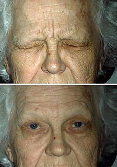
Picture 6: The upper photo depicts essential blepharospasm. The lower photo was taken after botulinum injection was administered to the orbicularis oculi of the patient.
Image Source: oculist.net
At the earlier course of the disease, the patient blinks frequently. It progresses in the form of uncontrolled spasms on one side of the eye. It worsens until both eyes are already affected [6].
Orbicularis Oculi Exercises

Picture 7: Relieving the orbicularis oculi of tension
Image Source: Kiviluoma L, Vital Face: Facial Exercises and Massage for Health and Beauty, Singing Dragon 2013, p 125
This is an exercise that will relax the muscle that closes your eyes. Stretch the orbicularis oculi with your fingers pulling against each other, as seen in the left photo of Picture 7. Below your eyebrows, massage the muscle in a gliding manner as seen in the right photo [7].
To increase the muscle tone around your eyes, do isometric exercises everyday for 10 minutes. Do this upright with your head straight. Look up without moving your head and “nearly” close your eyes. You will feel the orbicularis oculi doing the act. Muscle tension caused by this exercise tones up your muscle [8].
References:
- Tortora GJ & Derrickson B, Principles of Anatomy and Physiology 12th edition, John Wiley & Sons Inc. 2009, pp 347-349
- Cunningham DJ, Cunningham’s Textbook of Anatomy, W. Wood 1818, p 450
- Stone RJ & Stone JA, Atlas of Skeletal Muscles 3rd edition, p 29
- Esmaeli B, Ophthalmic Oncology, Springer 2011, p 299
- Hansen JT, Netter’s Clinical Anatomy 2nd edition, Saunders 2010, p 385
- Newman NJ et al, Walsh and Hoyt’s Clinical Neuro-Ophthalmology, Lippincott Williams & Wilkins 2008, p 491-491
- Kiviluoma L, Vital Face: Facial Exercises and Massage for Health and Beauty, Singing Dragon 2013, p 125
- Copley A, How to Get Rid of Hollows Under the Eyes Naturally, Without Surgery, Medical Bills or Cosmetics accessed on http://voices.yahoo.com/how-rid-hollows-under-eyes-naturally-6818584.html